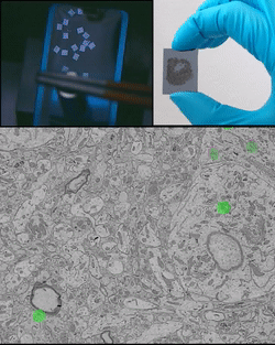
The animation on the top left shows remote magnetic actuation of ultrathin sections of biological tissue.
The image on the top right shows a piece of silicon wafer containing 1015 consecutive ultrathin sections ready for correlative light and electron microscopy imaging.
The animation on the bottom shows consecutive ultrathin sections acquired with multichannel fluorescent and electron microscopy. The color puncta show the locations of two different types of neuroanatomical tracers.
Large scale correlative light and electron microscopy for neural circuit reconstruction
Observing all neurons, their fibers and synapses in a small volume of brain tissue can only be performed with electron microscopy in volumes typically smaller than a tenth of a millimeter cube. Considering a piece of neuron in such a small excised volume, how to assess where in the brain does it receive inputs from and where does it send outputs to ?
We are solving this challenge with a combination of new developments in sample preparation and imaging:
- We are developing a new sample preparation technology for the collection of ultrathin sections on substrates suitable for high throughput and correlative light and electron microscopy.
- We are further improving sample preparation protocols along with establishing a library of neuroanatomical tracers suitable for correlative light and eletron microscopy imaging. It will allow us to visualize the location of neuroanatomical tracer molecules in electron microscopy imagery, thereby determining neuron projection types, that is, where do neurons receive input from and where do they send input to.
- We are developing an automated imaging pipeline for the acquisition and assembly of large correlative light and electron microscopy datasets.